Presurgical Planning
 Sometimes it is very important to know which brain functions are in the neighborhood of a lesion, particularly if that lesion is going to be removed by surgery. In lesions formed before birth, such as blood vessel abnormalities and some tumors, the areas of language and other mental process can be misplaced. fMRI shows the actual location of the functions and their spatial relationship with those lesions.
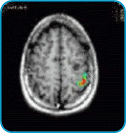
In the left image there is a little cyst in the left hemisphere (seen at the right side of the brain as a round black shape). This lesion produced seizures. The surgeon needs to know the location of the cortex in charge of the movement of the hand. This fMRI shows in colors the area used for the movement of the right fingers in a tapping task. This enables the neurosurgeon to extract the lesion without removing the area needed to move the hand.
Language Mapping
Brain functions can be divided into basic and complex. Basic functions require few areas of the cortex, while complex functions could be spread over one hemisphere, or even in both. Basic brain functions are consistently represented in the same places across different people, while the complex functions tend to vary from person to person. Language is a typical complex function. In most people, language is represented in the left hemisphere, but it is completely normal to find representation in the right hemisphere or divided between them. Prior to brain surgery, it is quite important to know the side in which the language is represented.
In epilepsy surgery this has a crucial importance. Some times it is necessary to remove part of the temporal lobe involved in the generation of seizures. The extension of the surgery must be restricted if it is found that this lobe is involved in language processing.
Here at Nicklaus Children's Hospital we have started to perform Language Mapping with fMRI to assess the localization of language, in adults and children over 7 years old.
Non-Surgical Applications
 fMRI has a great potential to provide insight into neurologic and psychiatric disorders. fMRI has been used to assess childhood stroke, migraine and epilepsy. This procedure has also been applied to children with developmental disorders, including autism, dyslexia, and other learning disabilities. Currently we are conducting research in sedated children and infants. Our goal is to obtain activation of the brain regardless of the sedation.
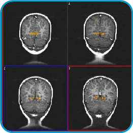
These images are from a 2 year-old child. They are named coronal cuts. The head is seen on top and the neck is on the bottom. Inside you see the two hemishpheres of the brain, and below them the cerebellum. The patient was sedated for a regular procedure. Flashing lights were presented during 3 minutes. The visual cortex, located in the occipital lobes, is depicted in red and yellow colors.
Images of activation in sedated kids have been obtained by presenting sounds (such as the mother's voice), or tactile stimulation to the skin (such as rubbing the hand).
On of the future purposes of fMRI is to predict developmental disorders at very young ages in order to provide effective early treatment interventions.